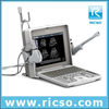
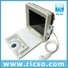
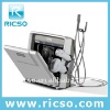
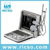
1) Image mode:B,2B,4B,B+M 2) Windwos XP embedded system 3) Memory disk:>160G 4) 12.1inch LED Look forward to Distributor
The latest model in 2012: Vetar V1
1, Memory disk: 4G(can support 32G)
2, Monitor: 12inch LCD
3, Probe connector: 2
1,General Descriptions
Image mode: | B,B+B,4B,B+M |
Scanning Depth: | ≥200mm (depend on the probe types) |
Resolution: | Lateral resolution≤2 mm Longitudinal resolution≤1mm |
Blind area: | ≤3mm |
Gray scale: | 256 |
Beam-forming: | - Full digital beam-forming technology(DBF) - Real-time Dynamic Aperture(RDA) - Dynamic Receiving Apodization(DRA) - Dynamic Receiving Focusing(DRF) - Dynamic Frequency Scan(DFS) - Tissue Harmonic Imaging (THI) |
2, Image processing
Gray-scale suppression
Gray mapping
Scanning angle selection
Γ-correction
Left-right reverse
Up-down reverse
Histogram
Edge Enhancement
Frame correlation
3,Functions
cine loop: | 256-frame cine loop memory ,playback speed selectable |
Memory disk: | 4G(can support 32G), |
Local zoom : | 2 times both in real-time and frozen |
OB measurement: | Equine,Bovine,Swine,Sheep,Cat,Dog |
E-Focus: | Number of focus,Focus distance,Focus position can be adjusted |
Monitor: | 12inch LCD |
Probe connector: | 2 |
Probes selectable: | convex, linear, micro-convex, rectal linear |
4,Peripheral Ports: PAL-D,RS-232,USB(2.0),VGA,LAN
5,Standard configurations: 5.0MHz Micro-Convex probe
6,Optional configuration: 6.5 Rectal Linear probe
Video Printer
Digital Portable VET Ultrasound Scanner


